Pathology
 The pathology and anatomy collections date back to the early nineteenth century, and relate not only to the University of Leeds and its predecessors (the Leeds School of Medicine and later the Yorkshire College of Science), but also the Leeds General Infirmary. They include biological specimens, wax models, microscope slides, illustrations and photographic glass plate slides. These were valuable resources for teaching, research and sharing knowledge amongst the professional pathology community.
The main areas are:
- A collection of historically important biological specimens displaying developmental abnormalities or effects of diseases which would be very unlikely to occur in such severe forms today. Please note that due to their sensitive nature, access to these specimens is restricted.
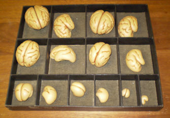
- A collection of wax models, primarily embryological. With real specimens both expensive and difficult to present, wax models were a more convenient way to provide a clear visual aid to teachers trying to explain the development of human and animal embryos as three-dimensional structures.
- The Matthew Stewart collection of slides, X-rays and photographic material, illustrations by Miss Ethel Wright, and related reports and correspondence. Matthew Stewart was Professor of Pathology at the University of Leeds between 1918 and 1951.